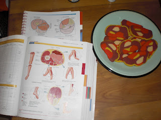
The other day I was in Anatomy lab looking at MRI images of the thigh, like this:
The Fellow who teaches my section jokingly described these axial views as "thigh cookies." From this I was inspired. Using a recipe for Pinwheel cookies often found at Christmas (http://allrecipes.com/recipe/
We made 2 red-based shades to make the different muscles, white for the bone, and yellow to make the nerves and skin.
Here's the part where I practice my anatomical knowledge, so feel free to skip ahead to the photos if you don't want to study with me. I started by forming the femur, complete with medial and lateral condyles. I then realized it would work best to build from the bottom up, so I formed the superficial posterior compartment, including the biceps femoris, semitendinosus, and semimembranosus. I then used the yellow dough to form a simplified version of the adductor canal, representing the saphenous nerve, femoral artery and femoral vein. I then placed the adductor muscles, including the adductor magnus which originates from the linea aspera on the femur. I added gracilis on the medial side, and then paused to take a picture.
You can see the condyles of the femur and the patella sticking out here. Then I popped that sucker in the fridge and went back to studying for a couple of hours.
When sufficiently cooled, I sliced up the thigh and set each cookie out to bake.
It looks pretty close to the real thing, if I do say so myself!
Then Hannah and I packed them up and gave a bunch to the Anatomy Fellows, a bunch to our classmates, and a huge pile to the 2nd year students who are taking their intimidating Neuro final as I type. Good luck, Class of 2015!


